
A rapid, targeted approach to detect chromosomal abnormalities with high specificity.
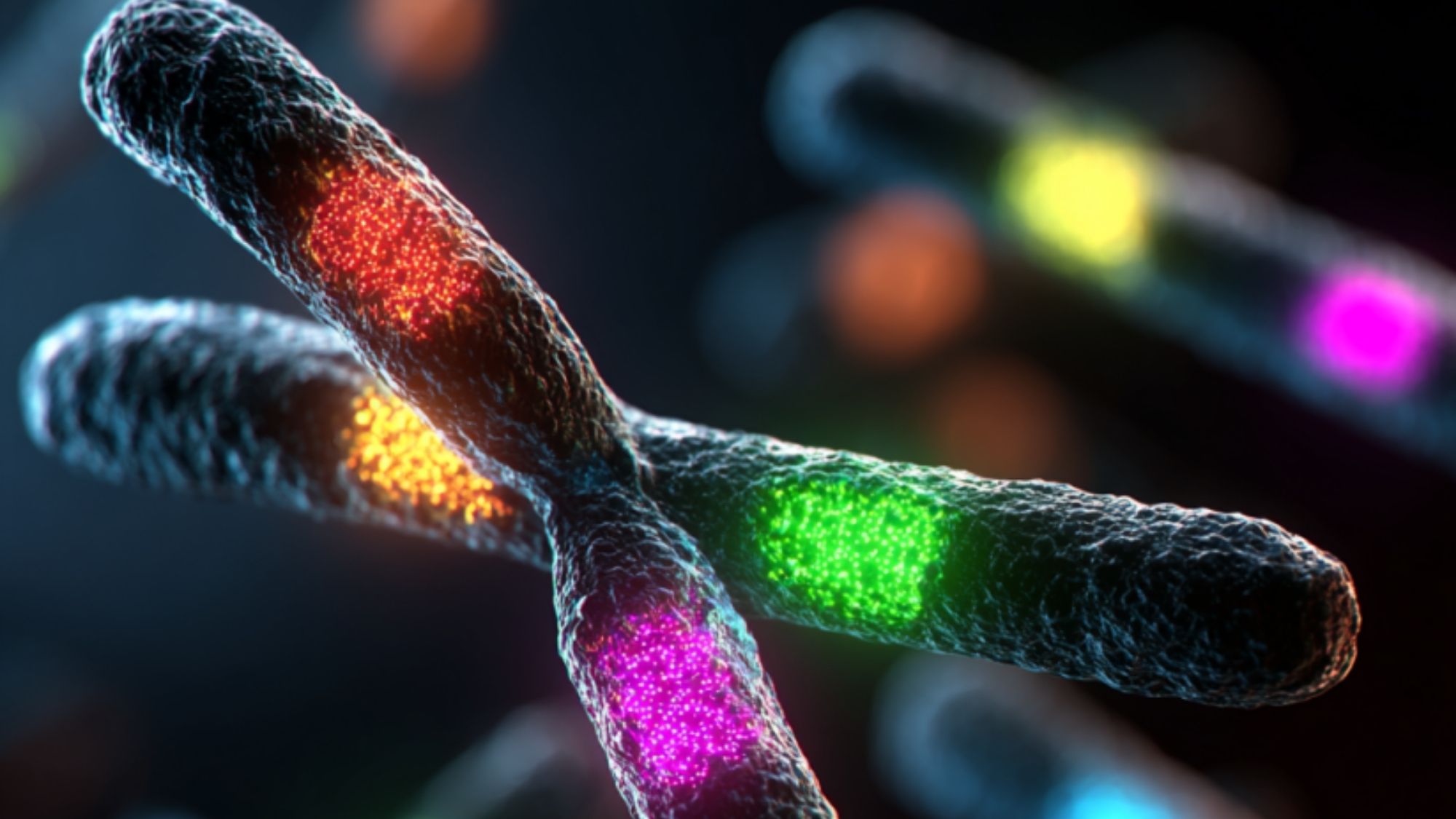
Fluorescence In Situ Hybridization (FISH) is a molecular cytogenetic technique used to detect and localize the presence or absence of specific DNA sequences on chromosomes. It uses fluorescent probes that bind to targeted regions, offering quick and precise analysis of chromosomal abnormalities—especially useful when time or specificity is critical.
Prenatal:
Postnatal:
Oncology:
Note: FISH is highly specific but limited to the targeted region being probed. It does not offer genome-wide screening.
Prenatal Samples
Postnatal Samples
Rapid Turnaround:
Targeted and Precise:
Complements Other Techniques:
Oncology & Prenatal Versatility:
Interpretable Reports:
